Did Biden ‘neglect’ Indonesia and ASEAN? The truth is darker: Global Times editorial
The 43rd ASEAN Summit and related meetings are scheduled from Monday to Thursday in Jakarta, Indonesia. Countries such as China, the US, Japan and South Korea will attend. According to Indonesian President Joko Widodo, "there are plenty [of agendas], but the focus is on the economy." This is a prominent feature of this year's ASEAN series of summits.
Continuously promoting regional economic development and win-win cooperation is a common aspiration of China and ASEAN. In this regard, there are many common languages. The pattern of China and ASEAN being each other's largest trading partners has been further consolidated. Immediately following the ASEAN Summit, the 20th China-ASEAN Expo and the China-ASEAN Business and Investment Summit will be held in Nanning, capital of China's Guangxi Zhuang Autonomous Region. These are significant and positive events for China and ASEAN's focus on regional economic cooperation.
Those who have some understanding of international events of the past two years know well that any multilateral occasion with participation of both China and the US tends to draw strong comparisons. This time is no exception.
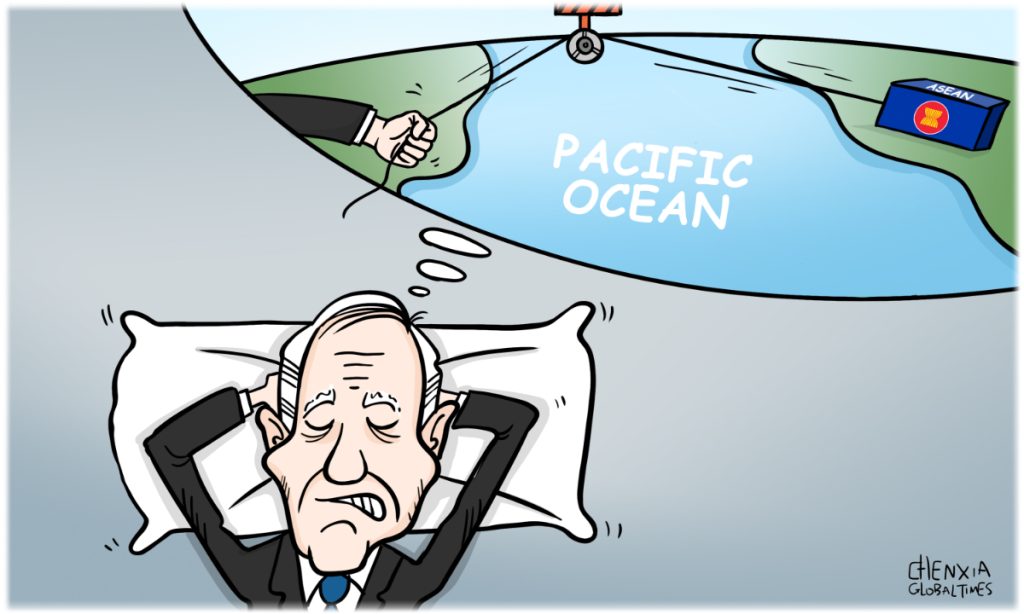
A few days ago, the US government announced that President Joe Biden will be absent from the summit and that Vice President Kamala Harris will attend in his place. Given that President Biden is scheduled to attend the G20 Summit in India shortly and then visit another ASEAN member, Vietnam, it gives the impression that Biden intentionally 'skipped' the ASEAN Summit hosted by Indonesia, which appears somewhat abrupt.
It is worth noting that various parties have different interpretations of Biden's absence, and the information revealed is rich and thought-provoking. As the host, Indonesia tactfully expressed disappointment at Biden's absence, which is understandable. Some held the view that "it is a signal to outgoing President Jokowi that Indonesia is simply not on the Administration's radar." Others say that this is the US' overall neglect of ASEAN and has "caused some frustration." The most widely circulated narrative in the US and Western media is that Biden's absence may cause Indonesia or ASEAN to "turn to China" or "find it difficult to resist China's maritime expansionary behavior." It can be seen that the mainstream perspective of the US in viewing ASEAN is to regard it as a sphere of influence it competes with China.
We believe that the Biden administration by no means intends to neglect Indonesia and ASEAN. On the contrary, it actually "takes them seriously"; but the real reason for attaching importance to Indonesia and ASEAN is not above board, and it is not consistent with ASEAN's actual interests and wishes, or even runs counter to them. Washington's lack of interest in the ASEAN Summit's focus on economic and trade cooperation is unequivocal.
Even if Biden attends the summit, it is conceivable that what he brings to the summit will be repeated geopolitical and security rhetoric, such as the so-called "freedom of navigation in the South China Sea" or "disputes over islands and reefs in the South China Sea." It will cause division on multilateral occasions and sow dissension, which is incompatible with the atmosphere of unity and cooperation at the summit. If these words are to be uttered by Harris, the negative impact may be relatively small. Therefore, Biden's absence is not a pity, even less of a loss for the ASEAN summit.
Why did Biden choose Vietnam over Indonesia? There are reasons. Since Washington did not do a good job in roping in ASEAN, it started to "split" it from within ASEAN. Additionally, Biden and Harris have been circling around China for the past year, so their intentions are quite obvious. It is easier for the US to pursue unilateral actions in bilateral settings than in multilateral platforms. But for ASEAN member countries that have been targeted by the US, it is necessary to keep an extra eye on the situation.
A recent article in the Los Angeles Times exposed Washington's true intentions. The article stated that while the Biden administration insists its approach "is not about forcing countries to choose, according to their interviews, "Washington has publicly and privately pressured ASEAN members to turn down China-proposed Belt and Road Initiative, reduce their economic and technological dependence on Beijing, and cancel their military partnerships with the People's Liberation Army. Does this not compel nations to choose sides? Washington's attempt to form a small clique is hurting rather than helping ASEAN's security concerns, according to even the US media.
Simply put, the US is currently the nation most difficult for ASEAN to deal with. There are more instances of it in the region, but the potential and practical advantages of development collaboration are dwindling as the risks rise. Every ASEAN meeting is essentially a testing ground for true and false multilateralism. It is not difficult to distinguish between those who come with genuine cooperation and win-win intentions and those who come with firewood and gasoline. ASEAN countries are well aware of this. If Washington picks the wrong course, the likelihood that it will fail increases along with the intensity of its frustration.